CSIRO unveils AI screening tech for early diagnosis of diabetic retinopathy

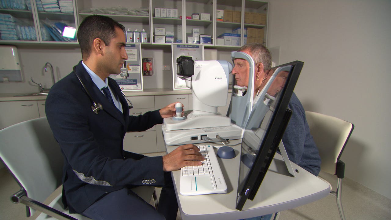
Dr Aly Khanbhai using the new scanning technology on a patient.
Australia's Commonwealth Scientific and Industrial Research Organisation (CSIRO) has announced that it has successfully trialled its artificial intelligence-powered eye-screening technology, Dr Grader, for the early diagnosis of diabetic retinopathy.
Dr Grader, developed in Perth, enables general practitioners to test patients for signs and identify the severity of the condition, which affects one in three diabetic patients in Australia and can lead to blindness.
According to the government-backed organisation, 1.7 million Australians are living with diabetes, and only specialists are able to screen for diabetic retinopathy.
"Patients at risk of this condition would usually be referred to a specialist for screening, waiting six weeks or more. Now it can potentially be done in a single 30-minute visit to a GP," professor Yogi Kanagasingam, the technology's creator and trial co-lead, said in a statement.
During the trial conducted at the GP Superclinic at Midland Railway Workshops in Perth, GPs screened 187 diabetic patients, taking high-resolution images of their eyes, which were then analysed by the technology for signs of the condition.
The images were also analysed by an ophthalmologist for comparison, and the technology was found to be as effective as a human specialist in detecting signs and grading its severity in patients, according to the CSIRO.
"Early detection and intervention for diabetic retinopathy is key, and this new tool is the first step to help GPs prioritise patients for treatment," Kanagasingam said.
"It could help avoid unnecessary referrals to public hospitals, potentially reduce waiting periods for patients, and enable ophthalmologists to focus on patients needing treatment and surgery.
Kanagasingam added that it could also help reduce the financial impact of diabetes on the Australian economy, estimated to be up to AU$14 billion a year.
The trial was funded through an NHMRC grant and base funding from WA Health and CSIRO through the Australian Telehealth Research and Development Group.
The software has been licensed by California-based TeleMedC, which develops ophthalmic diagnostic imaging systems for face-to-face and virtual medical consultations. The company will commercialise Dr Grader, with plans to install it at 20 GP clinics in Western Australia over the coming months before expanding it across Australia.
The CSIRO ran a teleophthalmology trial in the Torres Strait Islands and Western Australian goldfields a few years ago, where nurses were trained to take retinal images using a mobile device. Those images were then forwarded for reading by ophthalmologists in Perth or Brisbane.
In February, IBM announced that its researchers in Melbourne had trained a research version of Watson to recognise abnormalities in retina images that could assist doctors in the early detection of eye diseases such as glaucoma, nicknamed "the silent thief of sight" as many patients remain undiagnosed until irreversible vision loss occurs.
Commencing in 2015, the IBM researchers applied deep learning techniques and image analytics technology to 88,000 de-identified retina images accessed through EyePACS to analyse key anomalies of the eye and streamline some of the manual processes that doctors have to undertake when diagnosing eye diseases.
This includes distinguishing between left and right eye images, evaluating the quality of retina scans, and measuring the ratio of the optic cup to disc, which is one of the key indicators of glaucoma.
A few months later, the company announced it had uncovered a new method -- which combines convolutional neural networks and dictionary-based learning -- to classify the severity of a patient's diabetic retinopathy. The method was found to be capable of returning results within 20 seconds and with 86 percent accuracy.