New silicon probe a breakthrough to fight diseases (photos)

IBM scientists, based in Zurich, Switzerland, have developed a flexible microscopic probe made from silicon that will help researchers and pathologists take tissue samples for drug studies and disease diagnostics.
The probe is made up of a silicon microfluidic head with two microchannels. The head head re-aspirates the liquid that it injects into a patient preventing the spread and accumulation of the liquid on the surface, which can lead to overexposure.
Here's more from IBM's press release.
Source: IBM Research - Zurich via flickr
This post was originally published on Smartplanet.com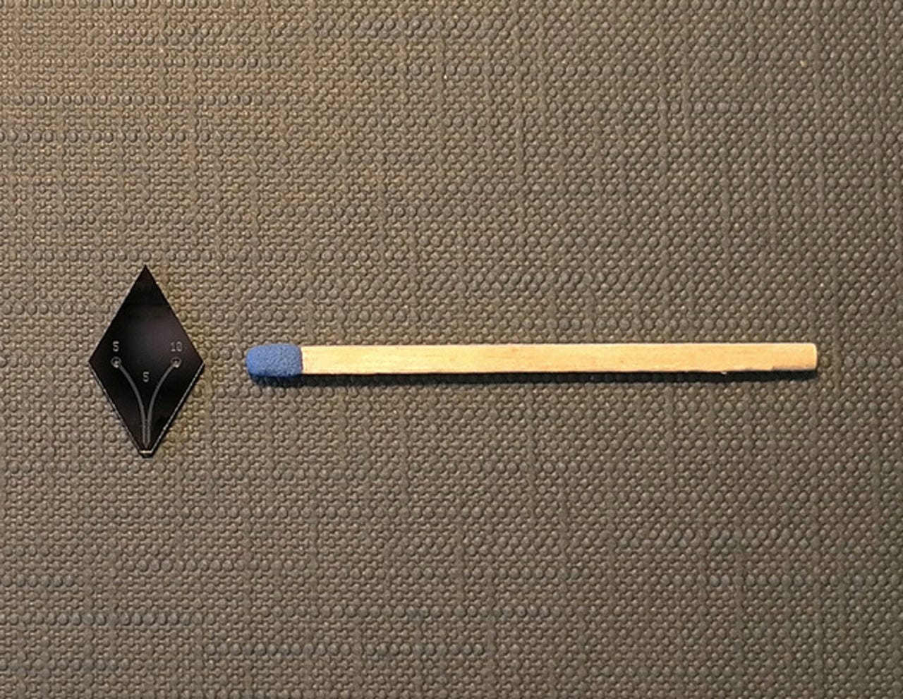
See how the probe compares in size with a matchstick.
Source: IBM Research - Zurich via flickr
This post was originally published on Smartplanet.comThe probe (see photo) consists of a silicon microfluidic head having two microchannels. Unlike an inkjet printer cartridge, the head re-aspirates the liquid that it injects on a surface. This prevents spreading and accumulation of the liquid on the surface, which can lead to overexposure.
Source: IBM Research - Zurich via flickr
This post was originally published on Smartplanet.comThe probe (see artistic rendering) consists of a silicon microfluidic head having two microchannels. Unlike an inkjet printer cartridge, the head re-aspirates the liquid that it injects on a surface. This prevents spreading and accumulation of the liquid on the surface, which can lead to overexposure.
Source: IBM Research - Zurich via flickr
This post was originally published on Smartplanet.comMicro- immunohistochemistry on cancerous breast tissue sections using different antibodies. (a) Monoplex staining of a 4-μm-thick well-differentiated invasive ductal carcinoma breast tissue section using a monoclonal mouse anti-human p53 protein antibody (54 μg ml-1, residence time 200 s). A line of hematoxylin is patterned with 1 s residence time for counterstaining. (b) Multiplexed staining of a well-differentiated invasive ductal carcinoma breast tissue section for the presence of p53 (α-p53: 54 μg ml-1, residence time of 300 s) and human progesterone receptor PR (α-PR: 125 μg ml-1, residence time of 300 s) with an additional hematoxylin counterstain. (c) μIHC processing of selected cores of an infiltrating ductal carcinoma breast tissue microarray, each 2 mm in diameter. Each core of the tissue array is characterized a priori by the supplier for the expression of ER, PR and p53. Four cores of the tissue microarray (locations A5, B5, B9 and C5) were processed with the vMFP (α-ER: 370 μg ml-1, α-PR: 125 μg ml-1 and α-p53: 54 μg ml-1).
Source: IBM Research - Zurich via flickr
This post was originally published on Smartplanet.comA concept and workflow of micro- immunohistochemistry (μIHC) using a vertical microfluidic probe (vMFP). Dewaxing and rehydration of the tissue are performed according to conventional IHC (1). Using injection and aspiration apertures at the apex of a vMFP head, a solution of primary antibody is hydrodynamically confined (in the presence of an immersion liquid) to selected areas of a tissue section (2). Post-processing for visualization of the antigens on the tissue section continues as in standard IHC: the tissue section is incubated with secondary antibodies, and enzymatic precipitation of 3,3'-diaminobenzidine (DAB) chromogen leads to a visual signal, indicating the expression level of specific antigens in the tissue section semi-quantitatively. Typical parameters for the vMFP scans are indicated.
Source: IBM Research - Zurich via flickr
This post was originally published on Smartplanet.comMicro- immunohistochemistry on cancerous breast tissue sections using different antibodies. (a) Monoplex staining of a 4-μm-thick well-differentiated invasive ductal carcinoma breast tissue section using a monoclonal mouse anti-human p53 protein antibody (54 μg ml-1, residence time 200 s). A line of hematoxylin is patterned with 1 s residence time for counterstaining.
Source: IBM Research - Zurich via flickr
This post was originally published on Smartplanet.comGovind Kaigala, a scientist at IBM Research - Zurich, prepares the microfluidic probe for testing.
Source: IBM Research - Zurich via flickr
This post was originally published on Smartplanet.comGovind Kaigala, a scientist at IBM Research - Zurich, looks at a stained tissue sample.
Source: IBM Research - Zurich via flickr
This post was originally published on Smartplanet.comMarios Georgiadis, currently a PhD student at ETH Zurich, Institute for Biomechanics, takes a closer look at a silicon wafer containing dozens of microfluidic probes.
Source: IBM Research - Zurich via flickr
This post was originally published on Smartplanet.comIBM scientist, Robert D. Lovchik, tests one of the microfluidic probes by blowing air through the micro channels.
Source: IBM Research - Zurich via flickr
This post was originally published on Smartplanet.comIBM scientist, Robert D. Lovchik, tests one of the microfluidic probes by blowing air through the micro channels.
Source: IBM Research - Zurich via flickr
This post was originally published on Smartplanet.comEmmanuel Delamarche, Experimental Biosciences, IBM Research - Zurich.
Source: IBM Research - Zurich via flickr
This post was originally published on Smartplanet.comThe team: top left, Robert D. Lovchik; Emmanuel Delamarche; bottom left, Govind V. Kaigala; and Marios Georgiadis.
Source: IBM Research - Zurich via flickr
This post was originally published on Smartplanet.com