Modded inkjet helps scientists study cell structures

Researchers at a US university have come up with a way of studying the mechanics of cells by printing them onto slides with a converted inkjet printer.
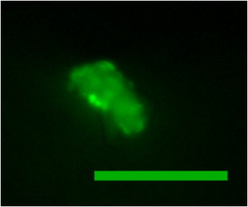
A printed cell is shown here with flourescent molecules inside. Image credit: Clemson University
The team used the temporary holes to inject fluorescent molecules that illuminated the cells, allowing them to see how the cell structures react to outside forces.
"We are actually interested in the cell mechanics of compressed cells. This method allows us to push on the cells and watch the response easily," the paper's author, Delphine Dean, said in a statement. "We are interested in cardiovascular cells, and how they respond to mechanical force."
According to Dean, the same technique can be used to print matrix proteins such as collagen onto substrates, for in vitro cell culture studies.
The Clemson team modified a standard HP DeskJet 500 printer by removing the paper feed mechanism and adding a slide feeder. The ink was also replaced with a cell solution.
"The advantage of using thermal ink-jet printing to inject molecules into cells is that the technique is relatively benign to cells," the team's paper read. "Cell viability after printing has been shown to be similar to standard cell plating methods. In addition, inkjet printing can process thousands of cells in minutes, which is much faster than manual microinjection."
One downside of the team's approach is that the temporary pores in the cells are only around 10nm in size, so only small proteins and particles can be injected. Also, printers cannot print in more than one dimension, so a specialised stage with adjustable height would be needed for layer-by-layer 3D bioprinting.