SNAC taps into Nvidia imaging tools to create AI algorithms for brain scan analyses

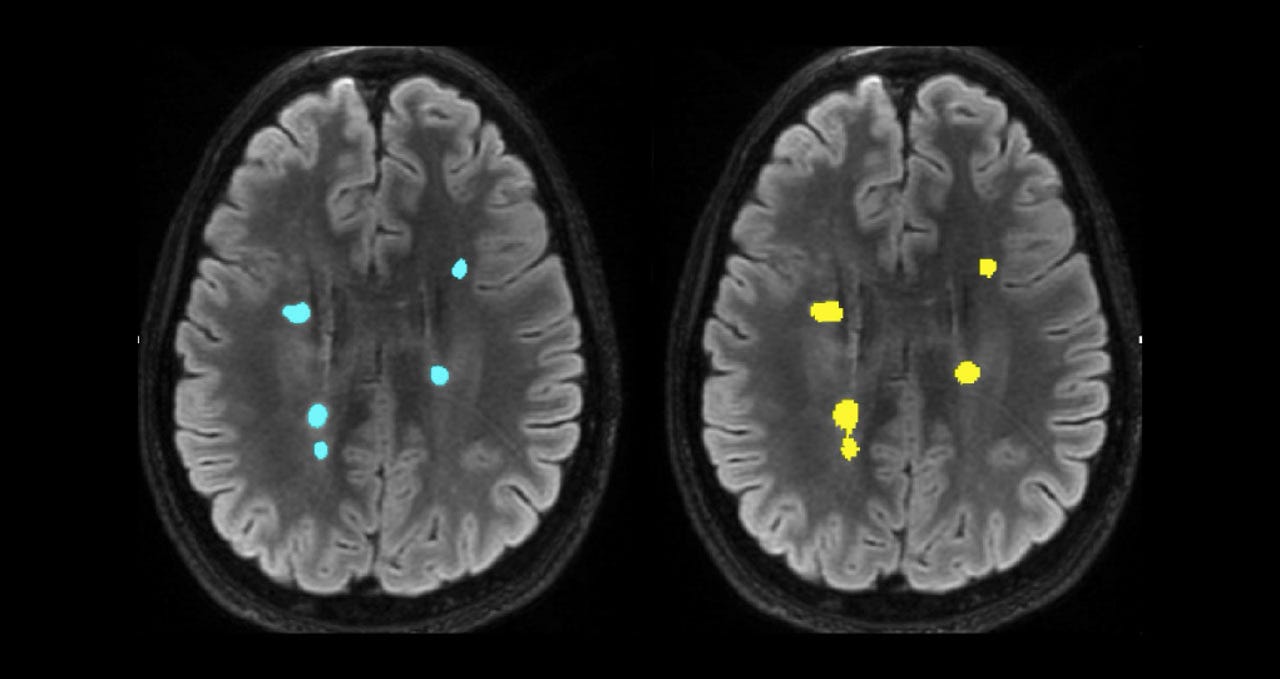
A side-by-side comparison of multiple sclerosis lesion segmentation. Left image shows manual lesion segmentation, while right shows fully automated lesion segmentation.
Sydney Neuroimaging Analysis Centre (SNAC) is building artificial intelligence (AI) tools to help radiologists automate laborious analysis tasks when it comes to examining brain scan imaging.
SNAC researchers have developed AI-based algorithms using the Nvidia Clara suite of medical imaging tools, as well as cuDNN libraries and TensorRT inference software for a number of applications, including isolating brain images from head scans and segmenting brain lesions.
According to neurologist professor at the University of Sydney's Brain and Mind Centre, Michael Barnett, within the one and a half years since SNAC integrated AI into its systems, it has helped "dramatically reduce the time taken to perform fairly straightforward structural assessments of MRI scans".
"For example, a prominent metric we want to produce for a clinical trial would be brain volume, lesion number and lesion volume, [and] change of lesions from one scan to another. Those processes would take up to an hour on a single scan, now we have 90-95% of the time fully automated," he said.
"Sure, with a clinical trial there is always a QA [quality assurance] step -- it's not like you can send scans off to pharmaceutical company without looking at it -- but nevertheless we're spending 2-3 minutes as opposed to 40 or 50 minutes.
"From a productivity and cost point of view from running a central reading service has been dramatic."
See also: Medical imaging at the 'speed of light': Nvidia's Clara supercomputer
Additional algorithms have also been developed to help radiologists examine progressive changes between longitudinal brain scans to see if patients are improving.
Typically, this process would involve scrolling up and down through 300 slices of brain images and checking back and forth between two scans placed side by side, Barnett said, but AI can now immediately highlight, segment, and quantitate those changes in a matter of seconds.
Barnett added that the algorithms are also applied to help look for new novel biomarkers in MRI and CT scans. Some immediate applications for this could include the detection of critical abnormalities on CT scans and rapidly bringing those scans to the attention of clinical radiologists or clinicians, he said.
Despite some immediate results, Barnett said we are only at the very beginning in terms of understanding the possibilities of what AI can do for imaging data.
"What we're doing is automating things to make it much easier and more productive. In two to three years it's going to totally change; and we're going start extracting information from scans that we never knew existed."
Solving the mysteries behind brain disease
One area that Barnett said he's been particularly interested in applying AI to is novel treatments of multiple sclerosis (MS), such as remyelination therapy and understanding how to assess the repair of nerves through brain scans.
"Most MS treatments target the immune system to reduce inflammation in the brain, but the remyelination therapies are the ones that will probably transform MS when it becomes available in the next five to 10 years," he said.
As a result, SNAC has now developed a remyelination biomarker platform underpinned by AI technology that will allow pharmaceutical companies to carry out clinical trials of emerging remyelinating therapies.
Similar work is already underway at SNAC for a cerebrovascular disease imaging biomarker.
"We're already applying some of our AI algorithms to that by working closely with The George Institute on a large multinational study called Trident," he said.
Beyond this, Barnett is optimistic the same work could be applied to other neurodegenerative diseases such as various dementias and eventually brain tumours.
Read also: How a companion robot can help children with chronic illness (TechRepublic)
Propelling imaging data with CRC funding
SNAC, which is co-located with the Brain and Mind Centre, has been able to expand its work in this space since being awarded close to AU$2.4 million last year through the federal government's Cooperative Research Centre (CRC) grant to integrate AI into the development of biomarkers and clinical neuroimaging.
As part of its research, SNAC works with imaging provider I-Med, the computational neuroscience team at the Brain and Mind Centre, as well as radiologists at Australian hospitals to validate its algorithms, giving SNAC access to curated datasets.
"Not a lot of people have curated data. Because we have this collaboration with I-Med, and we're housed in the Brain and Mind Centre and have a large number of clinics and we see a lot of patients, all of the data is very tightly curated, it means we have amazing ground truth for training our AI model," Barnett said.
Related Coverage
- How AI is making healthcare human again
- How machines are beating cardiologists in North Central Pennsylvania
- How industry cloud technology is changing healthcare
- Sydney healthcare clinicians turn to data analytics to improve back pain treatments
- Mary Meeker's 2019 Internet Trends report spotlights healthcare digitization
- CVS Health's grand consumer-driven healthcare plan depends on data, infrastructure
- AI in medicine: Tech that can improve the patient-doctor experience (TechRepublic)