Photos: Tech unlocks secrets of ancient Mummy

State of the art MRT scanner peers into history
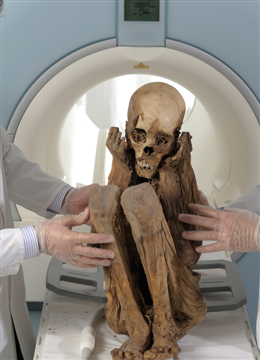
A millennium after his death this boy from Peru, pictured here, is revealing secrets to Swiss scientists thanks to pioneering new scanning technology. The magnetic resonance tomography (MRT) scanner and new analysis technique developed by Siemens Healthcare offered an unrivalled glimpse inside his mummified corpse.
Photo credit: Siemens
The mummy is loaded into the MRT scanner, which creates a strong magnetic field that causes hydrogen atoms within the corpse to generate radio waves. The scanner uses these waves to build a map of the atoms and generate an image of the tissue.
Photo credit: Siemens
The scan, being examined here by scientists at the University of Zurich, revealed the mummy's intervertebral disks, cerebral membrane, blood vessels, and embalming fluid residue. The ephyseal plates of the mummy’s upper arm were also clearly recognisable.
Photo credit: Siemens
Doctors have been using MRT for 25 years to generate precisely detailed 3D images of the human body, without the need to expose patients to X-rays. But the Ultra-short Echo Time measuring technique used here offers a snapshot of dry tissues, such as proteins and bones as shown here, that were previously impossible to image using MRT.
Photo credit: Siemens
The new diagnosis technique could be used in the future to examine metabolic processes in the heart and identify abnormal changes to the body’s metabolism or the brains of Alzheimer patients in scans such as the one pictured.
Photo credit: Siemens